
PATIENT EDUCATION
Epiretinal Membrane (Macular Pucker)
An epiretinal membrane is a thin layer of tissue on the macula that may cause blurred or distorted central vision. Many remain mild and can be monitored.
Sudden symptoms may have another cause
Epiretinal membranes usually change vision gradually. Seek prompt care for sudden flashes, a shower of floaters, a curtain or shadow, abrupt vision loss, or significant eye pain.
WHAT IS AN EPIRETINAL MEMBRANE?
A thin membrane can wrinkle the macula
The macula is the central part of the retina used for reading, recognizing faces, and seeing fine detail. An epiretinal membrane—also called a macular pucker—forms on the retinal surface. If it contracts, it can distort the macula and change central vision.
Most epiretinal membranes are related to normal age-related changes in the vitreous. They may also occur after a retinal tear or detachment, inflammation, trauma, vascular disease, or previous eye surgery.
SYMPTOMS
Vision changes range from none to significant distortion
Blurred central vision
Fine print, faces, and detailed tasks may appear less clear even with an updated glasses prescription.
Wavy or bent lines
Straight lines, doorframes, or text may look curved or uneven. This is called metamorphopsia.
Different image size
Objects may appear larger or smaller in one eye, making binocular vision uncomfortable.
Double or shadowed vision
A single eye may produce ghosting or doubled images when the membrane distorts the macula.
AT-HOME MONITORING
An Amsler grid can help track visual distortion
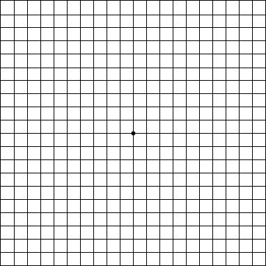
An Amsler grid is a simple home-monitoring tool. Viewed one eye at a time, it may make new waviness, missing areas, or changes in central vision easier to notice.
It does not replace an eye examination or OCT imaging. Contact your eye doctor if the grid looks newly or increasingly distorted.
DIAGNOSIS
OCT shows the membrane and retinal distortion
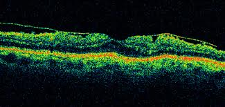
A dilated retinal examination may show a sheen or wrinkling over the macula. Optical coherence tomography (OCT) provides detailed cross-sectional images, confirming the membrane and showing retinal thickening, cystic changes, or traction.
Your clinician compares visual acuity, symptoms, OCT images, and changes over time to determine whether observation or treatment is appropriate.
OBSERVATION OR SURGERY
Treatment depends on how much the membrane affects daily life
Observation
Mild membranes with little distortion can often be monitored with examinations and OCT. Glasses may improve associated refractive blur but do not remove the membrane.
Vitrectomy and membrane peel
When blur or distortion is functionally significant, a retina surgeon may remove the vitreous gel and carefully peel the membrane from the macula.
Expected improvement
Vision often improves gradually over months after surgery, but the amount of improvement varies. Some distortion may remain, especially when the membrane has been present for a long time.
Risks and cataract progression
Surgery carries risks including infection, bleeding, retinal tear or detachment, and recurrence. Cataracts often progress after vitrectomy in patients who still have their natural lens.
COMMON QUESTIONS
Epiretinal membrane FAQ
Will it become a retinal detachment?
An epiretinal membrane is different from a retinal detachment. It sits on the retinal surface rather than separating the retina from the eye wall.
Do eye drops treat it?
No eye drop removes an epiretinal membrane. Treatment is observation or surgery depending on symptoms and retinal findings.
When should surgery be considered?
There is no single visual-acuity cutoff. The decision is based on distortion, functional limitations, OCT findings, and the expected benefit versus surgical risk.
Can it return after surgery?
Recurrence is possible but not common. Follow-up examinations monitor healing and any new membrane formation.
PATIENT EDUCATION VIDEO
Learn more about epiretinal membranes
This video provides general education and does not replace an eye examination or individualized medical advice.
THE EYE CENTERS
Noticing distortion in one eye?
A dilated examination and OCT can determine whether an epiretinal membrane or another macular condition is responsible.